Home

Piantare alberi Patois Terracotta acetone fixation for immunofluorescence Pratico sposa Dimora

Successful Immunofluorescence: Fixation and Permeabilization

An evaluation of fixation methods: Spatial and compositional cellular  changes observed by Raman imaging - ScienceDirect
An evaluation of fixation methods: Spatial and compositional cellular changes observed by Raman imaging - ScienceDirect

Immunocytochemistry Protocol

1.2 Fixation–Fixed cell imaging: 5 steps for publication-quality images -  YouTube
1.2 Fixation–Fixed cell imaging: 5 steps for publication-quality images - YouTube

Our 9 tips to optimize your immunofluorescence staining

Optimization of cell transfer and fixation for immunofluorescence... |  Download Scientific Diagram
Optimization of cell transfer and fixation for immunofluorescence... | Download Scientific Diagram

Key Tips on Sample Preparation for Immunofluorescence | Biocompare Bench  Tips
Key Tips on Sample Preparation for Immunofluorescence | Biocompare Bench Tips

Immunohistochemistry

Fixation and Permeabilization in ICC IF: Novus Biologicals

Immunolabeling artifacts and the need for live-cell imaging | Nature Methods

Immunofluorescence Guide - Creative Diagnostics

Fixation/Permeabilization: New Alternative Procedure for Immunofluorescence  and mRNA In Situ Hybridization of Vertebrate and Invertebrate Embryos -  Fernández - 2013 - Developmental Dynamics - Wiley Online Library
Fixation/Permeabilization: New Alternative Procedure for Immunofluorescence and mRNA In Situ Hybridization of Vertebrate and Invertebrate Embryos - Fernández - 2013 - Developmental Dynamics - Wiley Online Library

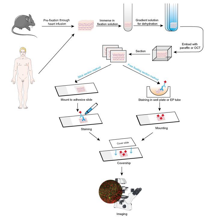
Tissue fixation, embedding and sectioning | Abcam

Immunofluorescence staining of paraformaldehyde-fixed HEp-2 cells... |  Download Scientific Diagram
Immunofluorescence staining of paraformaldehyde-fixed HEp-2 cells... | Download Scientific Diagram

Immunofluorescence staining of IFN-treated MG63 cells fixed with... |  Download Scientific Diagram
Immunofluorescence staining of IFN-treated MG63 cells fixed with... | Download Scientific Diagram

Immunofluorescence: Tips for Immunostaining Cultured Cells | Proteintech  Group
Immunofluorescence: Tips for Immunostaining Cultured Cells | Proteintech Group

Evaluation of TCA-fixation. (A) MTD-1A cells were fixed with 10% TCA (a...  | Download Scientific Diagram
Evaluation of TCA-fixation. (A) MTD-1A cells were fixed with 10% TCA (a... | Download Scientific Diagram

An evaluation of fixation methods: Spatial and compositional cellular  changes observed by Raman imaging - ScienceDirect
An evaluation of fixation methods: Spatial and compositional cellular changes observed by Raman imaging - ScienceDirect

Representative examples of immunofluorescence staining of mouse fixed... |  Download Scientific Diagram
Representative examples of immunofluorescence staining of mouse fixed... | Download Scientific Diagram

Representative examples of double immunofluorescence staining of... |  Download Scientific Diagram
Representative examples of double immunofluorescence staining of... | Download Scientific Diagram
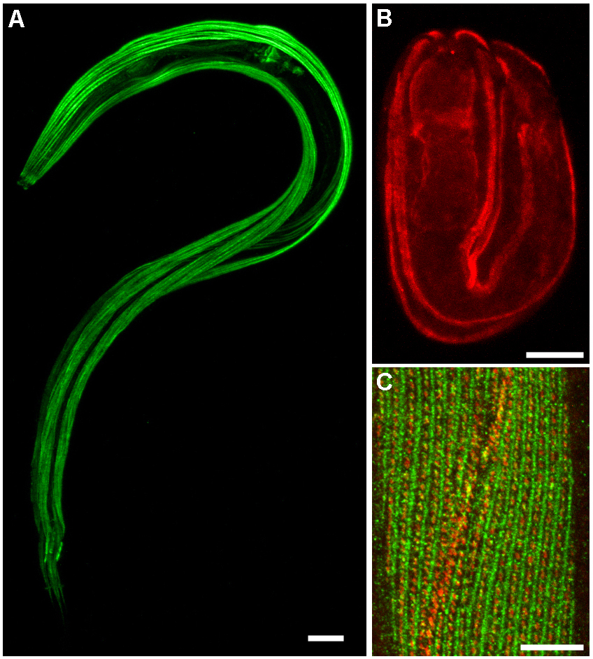
Immunohistochemistry

Fixing with formaldehyde and permeabilizing with acetone differs from... |  Download Scientific Diagram
Fixing with formaldehyde and permeabilizing with acetone differs from... | Download Scientific Diagram
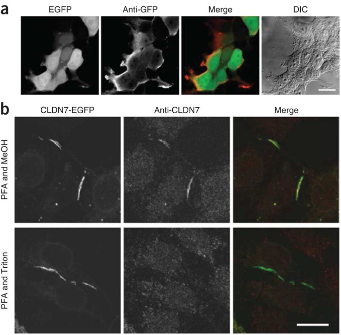
Immunofluorescence Protocol | StressMarq

Fixation and Permeabilization in ICC IF: Novus Biologicals

How to Prepare Your Specimen for Immunofluorescence Microscopy | Science  Lab | Leica Microsystems
How to Prepare Your Specimen for Immunofluorescence Microscopy | Science Lab | Leica Microsystems